
-
1
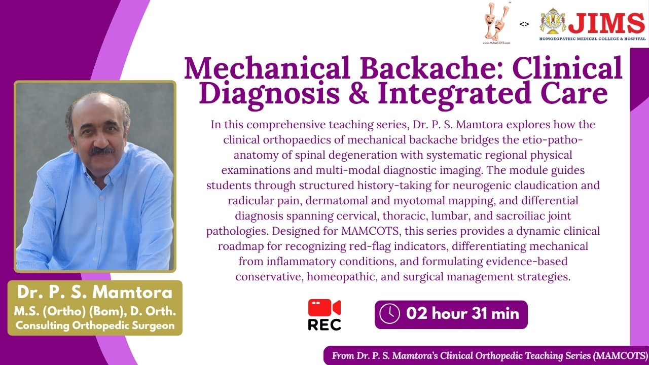
 Mechanical Backache - History Taking: Symptomatology + Neurology
Wide array of onset in Mechanical backache: acute, subacute, chronic or acute on chronic. Acute onset usually occurs in a younger age group, whereas the chronic variety is seen in the elderly; often, in both cases, backache worsens with activity and improves with rest. Associated neurological symptoms may include radicular pain along the dermatome following a specific activity, with or without paresthesiae, claudication, or motor weakness. Neurological examination of that particular dermatome may confirm the disc level before the MRI.
-
2
Mechanical Backache - History Taking: Claudication
Subacute or chronic backache due to canal stenosis often causes neurogenic claudication. Classic pointers toward neurogenic claudication include proximal involvement, associated backache, paresthesia, and pain relief with spinal flexion, unlike vascular claudication, which is more distal and relieved by rest. A saddle-shaped thrombus at the aortic bifurcation can mimic neurogenic claudication but is differentiated by absent distal pulses.
-
3
Mechanical Backache - History Taking: Nitty Gritty
Localised pain is common in mechanical backache, compared with ill-defined or generalised pain. Radiation of pain along a dermatome is specific to mechanical backache. However, causes can overlap, such as poor posture and mental stress, which can act as catalysts for early degenerative changes in the spine.
-
4
Mechanical Backache - Clinical Examination: Cervical Spine
The examination of the spine can be divided into cervical, dorsal and lumbar depending on the location of the symptom. Each can be examined using the following format: spinal signs (tenderness, asymmetric spasm, and asymmetry in range of motion); stretch signs to elicit nerve root compression; and neurological signs (motor and sensory).
-
5
Mechanical Backache - Clinical examination: Dorsal Spine
Examination of Dorsal spine begins with inspection (to look out for scoliosis, increased kyphosis) and check the range of motion - flexion and extension.
-
6
Mechanical Backache - Clinical Examination: Lumbar Spine
The examination of the spine is divided into Inspection (gait, increased curvature), Palpation (to feel for tenderness, spasm), Range of motion (flexion of the spine occurring at the hip or the lumbar spine), stretch tests to assess acute nerve root compression (SLR, femoral stretch test) and Neurological examination, sensory and motor along the dermatome. Movements of the spine that cause pain and asymmetry in range of motion belong to mechanical backache.
-
7
Mechanical Backache - Clinical Examination of Back: SI Joint
Active SLR causing pain at the SI joint, not felt during passive SLR, suggests SI joint pathology. A positive gluteal stretch test or SI joint stretch test (figure-of-4) that reproduces pain at the SI joint points to SI joint pathology. Tenderness at the SI joint or pain elicited by posterior-to-anterior compression of the sacrum are signs of SI joint pathology.
-
8
Mechanical Backache - Etio-patho-anatomy of Degeneration of Spine ( Stage1 )
Spinal degeneration consists of 3 stages; the progression and pace of degeneration may vary from vertebra to vertebra. Stage 1 represents dysfunction, which at the disc level is demonstrated as a radial or circumferential tear. The nucleus pulposus may herniate through the tear in the annulus, triggering a cascade of inflammatory mediators that irritate the sinuvertebral nerve and cause pain and muscle spasm.
-
9
Mechanical Backache - Etio-patho-anatomy of Degeneration of Spine ( Stage 2 )
With recurrent annular tears in the disc and inflammation at the facet joints, this results in instability due to hypermobility and capsular stretch. The clinical presentation may comprise pain at the affected level with certain movements. The disc changes from a gel-like, shock-absorbing substance to a desiccated one with reduced shock absorption, with the surrounding muscles bearing the brunt.
-
10
Mechanical Backache - Etio-patho-anatomy of Degeneration of Spine (Stage 3)
Phase 1 of spinal degeneration can present as pain and stiffness, which may be related to synovitis at the facet joints and microscopic disc tears, with loss of curvature due to muscle spasm. Stage 2 demonstrates capsular laxity and subluxation of the facet joints, and at the disc level, disc herniation progressing to disc extrusion and disc osteophytes leading to canal stenosis (stage 3.
-
11
Mechanical Backache - Etio-patho-anatomy of Degeneration of Spine (Visuals)
Degenerative disc disease may lead to disc collapse and a consequent tear in the annulus, resulting in a disc bulge, protrusion, extrusion, and sequestration. Loss of intervertebral space can narrow the neural foramina, which can compress the nerve roots. Disc degeneration and vertebral degeneration may go hand in hand with resultant osteophytes and bony spurs that can further compress the nerve root.
-
12
Mechanical Backache - Clinical Picture for Diagnosing Cervical & Lumbar Discogenic Pain
Clinical assessment of the myotomes and the dermatomes of the cervical and lumbar nerve roots to predict the level of the disc bulge/herniation causing the respective nerve root compression.
-
13
Mechanical Backache - Case Discussion
This video splendidly demonstrates the connection of the symptom to the pathology, anatomy and the likelihood of the corresponding diagnosis.
-
14
Mechanical Backache - Differentiation between Cervical Spine & Shoulder Joint Pathology
Affection of the C4-5 disc involving deltoid muscle weakness may present as shoulder pain. In shoulder pathology, shoulder range of movement is painfully restricted or affected, whereas in cervical pathology, shoulder range of movement is full, with only deltoid weakness. In cervical pathology, hanging the affected-side limb aggravates the pain, whereas raising the upper limb over the head relieves it; the reverse is true in shoulder pathology.
-
15
Mechanical Backache - Cervical Spine, Vertigo, Headache
-
16
Mechanical Backache - SI Joint Affection
Sacroiliac joint dysfunction, or Sacroiliac joint strain, is a mechanical, non-inflammatory condition that manifests as localised SI joint pain of acute origin and, on examination, is confirmed by a positive SI joint stress test, pelvic malalignment, and tenderness at the SI joint. Radiologically, condensans ilii is a differential diagnosis for sacroiliitis; however, it is benign, with a normal appearance and no irregularity, erosions, or joint space narrowing.
-
17
Mechanical Backache - Thoracic Outlet Syndrome
Radicular pain or paresthesia along the medial aspect of the little finger may be a presentation of thoracic outlet syndrome and is less likely a cervical disc pathology.
-
18
Mechanical Backache - Cervical Spine Degeneration X -Rays
A plain X-ray can never diagnose an acute lumbar or cervical disc prolapse, which is best assessed with a thorough clinical history and examination and confirmed by MRI. Loss of cervical lordosis on X-ray indicates paraspinal muscle spasm. Degenerative features on X-ray include anterior or posterior osteophytes, reduced disc space, ossification of the anterior longitudinal ligament, loss of cervical lordosis, and uncovertebral sclerosis, pointing toward uncovertebral arthropathy.
-
19
Mechanical Backache - Dorsal Spine Degeneration X-Rays
In an AP view of the dorsal spine X-ray, the pedicles are seen end-on. A pedicle that is not distinctly seen may indicate pathology or erosion.
-
20
Mechanical Backache - Lumbar Spine Degeneration X-Rays
The degenerative process in the lumbar spine is radiologically demonstrated by reduced disc space, narrowed intervertebral foramen, subchondral sclerosis, or osteophytes, which should be correlated clinically. The vacuum phenomenon appears as a radiolucent zone in the disc and is often seen in severe disc degeneration. The Scottie dog sign is appreciated on the oblique radiographs of the normal lumbar spine; however, in spondylolysis it may be seen as 'a stretched-out neck of the dog' due to a break in the pars interarticularis.
-
21
Mechanical Backache - MRI Findings
-
22
Mechanical Backache - Different Pathologies
Mechanical backache is an umbrella term that encompasses a range of spine affections such as degenerative spine pathologies - OA spine, facetal OA, degenerative disc disease, canal stenosis, spondylolisthesis and spondylolysis.
-
23
Mechanical Backache - Difference between Inflammatory v/s Mechanical
Characteristics of Inflammatory backache are - diffuse pain along the spine, worse by rest better by activity and may have associated extra articular manifestations of other systems (Eye, Skin, GUT, GIT ) in contrast to mechanical backaches, which are localized, better by rest and worse on activity and radiation of the pain is along the dermatome with or without associated neurological signs and symptoms.
-
24
Mechanical Backache - Disc prolapse Management
Management of Acute Disc Prolapse comprises rest, pharmacological treatment, and physiotherapy, all of which should begin as soon as possible.
-
25
Mechanical Backache - Disc Prolapse Management: Long Term Management
Physical therapy, along with medical treatment, is equally essential, and an optimum customised rehabilitation protocol must be part of the long-term management strategy. Equal emphasis on proper ergonomics and posture correction, along with appropriate exercises for an extended period, cannot be overstated. However, all treatment modalities are palliative and cannot reverse the disc bulge or the desiccated disc. However, it is imperative to prevent further relapses; one ought to diligently follow the rehabilitation program as advised.
-
26
Mechanical Backache - Disc Prolapse Management: Surgical Indications
Bladder-bowel involvement, as in Cauda Equina syndrome, and acute onset of paraparesis or quadriparesis with chronic backache mandate urgent decompression spine surgery. Highly intense backache recalcitrant to rest and medicines, even when accompanied by a mild neurodeficit, or recurrent backache episodes with neurodeficit, is reason enough for surgery.
-
27
Mechanical Backache - Different Stages of Degenerative Spine with Correlating Management
A acute disc prolapse treatment regime along with anti inflammatory and antispasmodic therapy incorporates rest for appropriate duration and avoidance of pain instigating movements. the key point to remember is that a desiccated disc can never have its hydrated status replenished and a disc bulge always predisposes the person for a subsequent episode anytime throughout his life. All the modalities involved in managing an acute mechanical backache are aimed at rehabilitating the patient to lead a functional, pain free life, however the structural changes cannot be reverted back.
-
28
Mechanical Backache - Cervical and Lumbar Spine Homoeopathic Specifics
Some Homoeopathic specifics - Left upper limb radiculopathy with periscapular pain may point towards KALMIA, severe unilateral neck pain with restricted range of motion, think of LACNANTHES, right lower limb pain with tight hamstrings and paresthesia - TELLURIUM, right lower limb pain with more of burning pain - GNAPHALIUM, canal stenosis with claudication and burning paresthesia -SULPHUR.
-
29
Mechanical Backache - Follow-up
Follow-up criteria for analysing disease progression are based on a detailed evaluation of symptomatology, comparison with previous presentation, and correlation with examination findings such as neurological deficits, SLR, and range of movement.
-
30
Mechanical Backache - Belts, Braces & Back Pain Myths
LS frame-type belts are generally recommended for spine instability seen in spondylolisthesis, though some belts can be used during acute, severe back pain until it subsides; long-term use of belts and braces is not advised. Backache following spinal anaesthesia is a fallacy, unless the needle has punctured the disc, causing pyogenic discitis.
-
31
 Feedback Form